Radio Diagnosis
All Departments
Low Vision Clinic of chirayu medical college and hospital helps the patients of Low Vision through a comprehensive rehabilitative process. By using specialized optics, teaching a patient to see around the damaged part of the visual system and developing a better localization patterns, patients learn to make the best use of their remaining vision, enabling many to return to the daily routines disrupted by vision loss.
SERVICES AVAILABLE
X-RAY MACHINES
The department equipped with Digital Radiography X-Ray Machine 800 mA
The digital format allows manipulation and magnification of the images to improve contrast and make the lesions look more conspicuous. The improved quality of images can almost eliminate the need for repeat radiographs.
Fluoroscopy with IITV X-Ray Machine 800 mA
600 mA X- RAY MACHINES
300mA X –RAY MACHINES AND mobile X-ray machine
OPG machine
Specialized contrast procedures such as Barium studies, intravenous Urography. Cystourethrograms , HSG are performed on these x-ray machines. The image intensifier TV systems used for performing these procedures enable more accurate diagnosis and significant reduction in radiation dose to patients.
MAMMOGRAPHY MACHINE
Breast imaging is performed with technology like mammography , breast ultrasound and breast MRI.We work with pathology team to perform breast biopsies as well.
Mammography and ultrasound guided needle biopsies and hook-wire localization procedures are performed in order to achieve a definitive diagnosis in nonpalpable cases of breast cancer.
USG MACHINES
The ultrasound facility of the department consists of the latest state of the art ultrasound scanners
Dimensional (2D) Ultrasound
» Abdomen
» KUB
» Pelvis
Small Parts Ultrasound
» Thyroid
» Breast (Sonomammogram)
» Superficial Swelling
» Scrotum & Eye (Ocular)
» Thorax (chest) Ultrasound
» Transcranial (Pediatric Neurosonogram) Ultrasound
» Transvaginal (3D) Ultrasound
» Neonatal and Paediatric Ultrasound
» Musculoskeletal Ultrasound
» Peripheral Nerve and Joint Ultrasound
DOPPLER STUDY
» Carotid and Vertebral Studies
» Portal Venous System
» Aorta and IVC Flow
» Arterial-Venous Systems
» Renal Doppler
» Transplant Kidney, Liver Doppler
» Illiac Vessels Doppler
» Obstetric Doppler
ASPIRATIONS
» Pleural
» Ascitis
» Pseudo cyst
» Abscess
» Pelvis Collection
» Ultrasound guided pigtail insertion
Image Guided Biopsy Using ultrasound & CT guided our highly skilled radiologists perform biopsies of the most intricate and complex organs including:
» Lung
» Liver
» Kidney
» Breast
» Musculoskeletal
» Retroperitoneum
» Pelvis
CT IMAGING
The Department has two CT scanners. A 16-slice Multi-detector CT & 16 slice CT with PET Scan . Procedures done using CT unit include:
» Whole body Imaging
» CT Angiography (Aorta, peripheral and cerebral)
» Bowel Imaging
» High Resolution Computed Tomography
» CT guided biopsy
» CT guided drainage procedures
» 3D Imaging Multiplanar Reconstruction including bronchoscopy
» Transplant imaging
Organ specific protocols for each imaging technique help achieve targeted and highly detailed examinations. The volume data of the CT images are processed in ADW 4.4 and EBW 4 workstations to produce excellent 3D reconstruction images.
MRI IMAGING
Magnetic Resonance Imaging of the whole body is performed in our department on the GE Signa 1.5 Tesla. The services using MRI include:
» Whole body MRI
» High Resolution Imaging of Joints
» MR Angiography
» MR Spectroscopy
» Perfusion and Diffusion Imaging
» Whole Spine Imaging
» MR Mammography
» Advanced abdominal imaging
» Whole Body STIR imaging for bony metastasis screening.
» Pre-operative MRI imaging for cochlear transplant.
INTERVENTIONAL RADIOLOGY
The Interventional Radiology (IR) Unit of the department provides both diagnostic and therapeutic services by using advanced imaging technology. The patients are treated with minimally invasive for early recovery and better quality of life of the patients.
FACILITIES AVAILABLE
» DSA
» Bronchial artery embolization
» Peripheral artery stenting
» PTBT
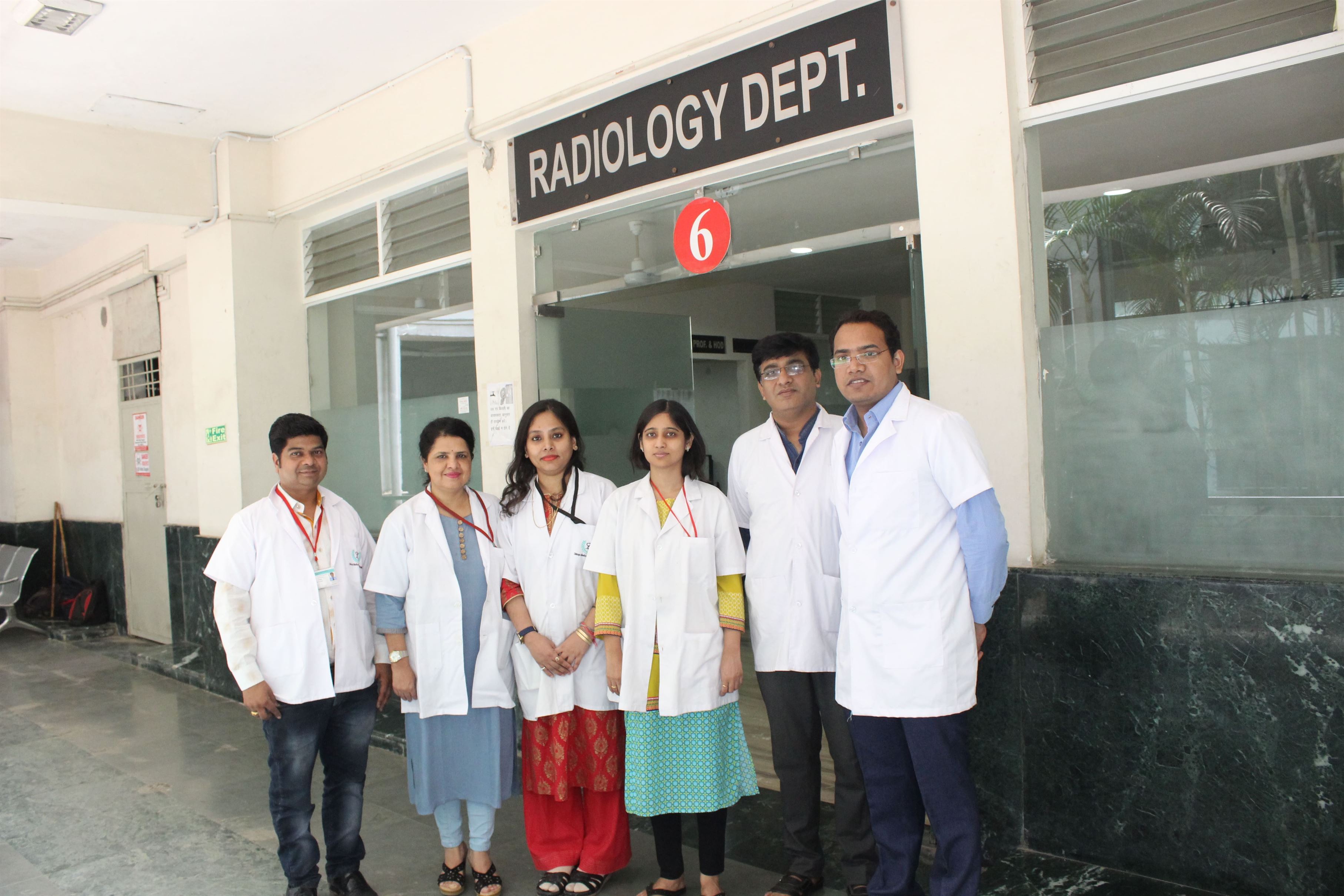
OUR DOCTERS TEAM


